Hepatocellular carcinoma 1
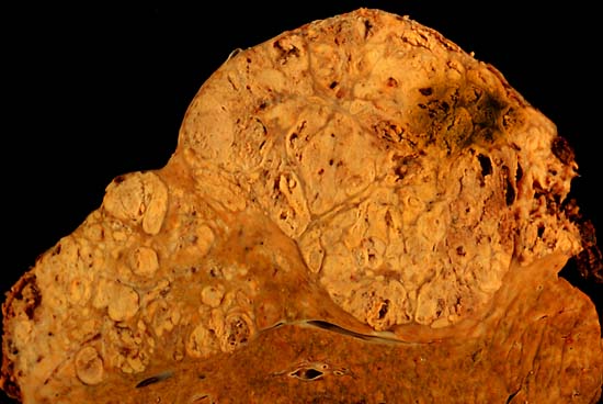
Hepatocellular carcinoma
This specimen is from a 50ish woman who presented to the hospital with abdominal pain and ascites. The radiologist recovered what appeared to be whole blood on paracentesis. Cytological exam of the bloody fluid showed no evidence of malignancy. Liver function tests were abnormal, and serologic tests were positive for antibody to hepatitis C. The patient deteriorated rapidly and died within a few days. The autopsy showed this hepatocellular carcinoma occupying much of the volume of a cirrhotic liver. Furthermore, the tumor had invaded the diaphragm and ruptured into the peritoneal cavity, causing the bloody ascites.
The photo shows a view of a longitudinal slice taken through the full length of the liver.
The photos were shot with a Minolta X-370 with 100 mm bellows lens on Kodak Elite ISO 100 transparency film. The specimen was sliced fresh and fixed in formalin overnight, then briefly immersed in 70% alcohol to retrieve some of the native color and dull the surface reflections.
Photograph by Ed Uthman, MD. Public domain. Posted 23 Sep 00Więcej informacji o licencji można znaleźć tutaj. Ostatnia aktualizacja: Fri, 16 Sep 2022 03:34:32 GMT