Śródmózgowie
| Ten artykuł od 2014-01 zawiera treści, przy których brakuje odnośników do źródeł. |
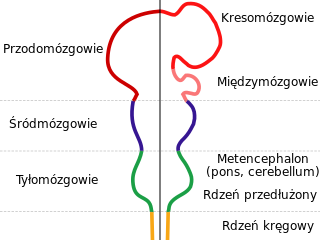
Po prawej pęcherzyki mózgowe pierwotne, a po lewej wtórne.

1. Wzgórki czworacze.
2. Wodociąg mózgu.
3. Istota szara środkowa.
4. Przestrzeń międzyodnogowa.
5. Bruzda boczna.
6. Istota czarna.
7. Jądro czerwienne nakrywki.
8. Nerw okoruchowy.
a. Wstęga (na niebiesko) z a’ wstęgą przyśrodkową i a" – wstęgą boczną.
b. Pęczek podłużny przyśrodkowy.
c. Szew mózgu.
d. Włókna skroniowo-mostowe.
e. Część wstęgi przyśrodkowej, która biegnie do jądra soczewkowatego i wyspy.
f. Włókna mózgowo-rdzeniowe (piramidowe).
g. Włókna czołowo-mostowe
Śródmózgowie (łac. mesencephalon) – środkowa część mózgowia u kręgowców, w której znajduje się tzw. wodociąg mózgu (aquaeductus cerebri) zwany też wodociągiem Sylwiusza łączący III i IV komorę mózgu. Śródmózgowie łączy się z móżdżkiem i rdzeniem przedłużonym oraz z międzymózgowiem. U ssaków część grzbietowa śródmózgowia utworzona jest przez blaszkę czworaczą (lamina quadrigemina), w której wyróżnia się wzgórki górne i dolne lub pokrywę wzrokową zróżnicowaną na ciałka bliźniacze (corpora bigemina), czyli płaty wzrokowe (lobi optici) u pozostałych kręgowców.
Śródmózgowie jest ośrodkiem wzrokowym, a u niższych kręgowców (ryby, płazy) jest także ośrodkiem integracji bodźców zmysłowych. U ssaków jest tylko ośrodkiem odruchowym zmysłów wzroku i słuchu.
Budowa
Śródmózgowie złożone jest z pokrywy śródmózgowia (tectum mesencephali) i konarów mózgu (pedunculi cerebri) (nakrywka (tegmentum) i odnogi mózgu (crura cerebri) współtworzą konary mózgu)[1]. Na przekroju poprzecznym płaszczyzna pozioma poprowadzona przez wodociąg mózgu jest granicą między pokrywą a nakrywką. Granicę między nakrywką a odnogą mózgu stanowią bruzdy: przyśrodkowa i boczna odnogi mózgu (sulcus medialis et lateralis cruris cerebri) oraz istota czarna (substantia nigra). Nakrywka i odnoga mózgu łącznie nazywa się konarem mózgu (pedunculus cerebri).
Pokrywa
Pokrywa śródmózgowia składa się przede wszystkim z blaszki pokrywy (lamina tecti), która zbudowana jest z:
- wzgórków górnych (colliculi superiores) – składają się z trzech warstw istoty szarej i czterech istoty białej; pełnią rolę ośrodka odruchów wzrokowych,
- wzgórków dolnych (colliculi inferiores) – pełnią rolę odruchowego ośrodka słuchowego,
- okolicy przedpokrywowej (regio pretectalis) – zawiera drobne jądra przedpokrywowe, do których dochodzi część łuku odruchu źrenicy na światło.
Nakrywka
Istotę szarą nakrywki śródmózgowia reprezentują jądra własne oraz jądra nerwów czaszkowych.
- Do jąder własnych należą:
- istota czarna (substantia nigra),
- istota szara środkowa (substantia grisea centralis),
- jądra tworu siatkowatego (nuclei formationis reticularis),
- jądro czerwienne (nucleus ruber).
- Do jąder nerwów czaszkowych należą:
- jądro ruchowe nerwu bloczkowego (nucleus motorius nervi trochlearis),
- jądro śródmózgowiowe nerwu trójdzielnego (nucleus mesencephalicus nervi trigemini),
- jądro główne nerwu okoruchowego (nucleus principalis nervi oculomotorii),
- jądro środkowe nerwu okoruchowego (nucleus centralis nervi oculomotorii seu Perli),
- jądro ogonowe środkowe nerwu okoruchowego (jądro Tschidy, nucleus caudalis centralis nervi oculomotorii seu Tschidi),
- jądro dodatkowe (autonomiczne) nerwu okoruchowego (nucleus accessorius seu autonomicus nervi oculomotorii seu Westphal-Edinegeri).
Istota biała nakrywki utworzona jest przez włókna:
- wstęgi przyśrodkowej,
- wstęgi bocznej,
- drogi rdzeniowo-pokrywowej,
- drogi móżdżkowo-czerwiennej,
- drogi rdzeniowo-siatkowej,
- drogi móżdżkowo-wzgórzowej,
- drogi pokrywowo-rdzeniowej,
- drogi czerwienno-rdzeniowej,
- drogi siatkowo-rdzeniowej,
- drogi pokrywowo-jądrowej,
- drogi czerwienno-jądrowej,
- drogi siatkowo-jądrowej,
- drogi suteczkowo-międzykonarowej,
- drogi uzdeczkowo-międzykonarowej,
- pęczka podłużnego przyśrodkowego,
- pęczka podłużnego grzbietowego.
Odnoga mózgu
Odnoga mózgu jest parzysta i zbudowana jest w całości z istoty białej, czyli włókien nerwowych przechodzących z torebki wewnętrznej do części brzusznej mostu.
Przypisy
- ↑ Adam Bochenek, Michał Reicher: Anatomia Człowieka Tom IV. Wydawnictwo Lekarskie PZWL, 2014. ISBN 978-83-200-4203-0.
Bibliografia
- Adam Krechowiecki, Florian Czerwiński: Zarys anatomii człowieka. Szczecin: Wydawnictwo Lekarskie PZWL, 2004. ISBN 83-200-3362-4.
- Śródmózgowie. W: J.Walocha, T.Iskra, J.Gorczyca, J.Zawiliński, J.Skrzat: Anatomia prawidłowa człowieka. Ośrodkowy układ nerwowy. Pod redakcją Andrzeja Skawiny. Kraków: Wydawnictwo Uniwersytetu Jagiellońskiego, 2003. ISBN 83-233-1756-9.
![]() Przeczytaj ostrzeżenie dotyczące informacji medycznych i pokrewnych zamieszczonych w Wikipedii.
Przeczytaj ostrzeżenie dotyczące informacji medycznych i pokrewnych zamieszczonych w Wikipedii.
Media użyte na tej stronie
The Star of Life, medical symbol used on some ambulances.
Star of Life was designed/created by a National Highway Traffic Safety Administration (US Gov) employee and is thus in the public domain.Star of life, blue version. Represents the Rod of Asclepius, with a snake around it, on a 6-branch star shaped as the cross of 3 thick 3:1 rectangles.
Design:
The logo is basically unicolor, most often a slate or medium blue, but this design uses a slightly lighter shade of blue for the outer outline of the cross, and the outlines of the rod and of the snake. The background is transparent (but the star includes a small inner plain white outline). This makes this image usable and visible on any background, including blue. The light shade of color for the outlines makes the form more visible at smaller resolutions, so that the image can easily be used as an icon.
This SVG file was manually created to specify alignments, to use only integers at the core 192x192 size, to get smooth curves on connection points (without any angle), to make a perfect logo centered in a exact square, to use a more precise geometry for the star and to use slate blue color with slightly lighter outlines on the cross, the rod and snake.
Finally, the SVG file is clean and contains no unnecessary XML elements or attributes, CSS styles or transforms that are usually added silently by common SVG editors (like Sodipodi or Inkscape) and that just pollute the final document, so it just needs the core SVG elements for the rendering. This is why its file size is so small.(c) I, Nrets, CC-BY-SA-3.0
Diagram depicting the main subdivisions of the embryonic vertebrate brain. The neural tube differentiates into forebrain, midbrain and hindbrain structures.
Axial section through mid-brain. (Schematic.) (Testut.) 1. Corpora quadrigemina. 2. Cerebral aqueduct. 3. Central gray stratum. 4. Interpeduncular space. 5. Sulcus lateralis. 6. Substantia nigra. 7. Red nucleus of tegmentum. 8. Oculomotor nerve, with 8’, its nucleus of origin. a. Lemniscus (in blue) with a’ the medial lemniscus and a" the lateral lemniscus. b. Medial longitudinal fasciculus. c. Raphé. d. Temporopontine fibers. e. Portion of medial lemniscus, which runs to the lentiform nucleus and insula. f. Cerebrospinal fibers. g. Frontopontine fibers.
Autor:
John A Beal, PhD
Dep't. of Cellular Biology & Anatomy, Louisiana State University Health Sciences Center Shreveport, Licencja: CC BY 2.5Human brainstem and thalamus - posterior view
- Taenia choroidea (and lateral: Lamina affixa, Stria terminalis)
- Thalamus, Pulvinar thalami
- (Ventriculus tertius)
- Stalk of Glandula pinealis
- Habenula
- Stria medullaris
- Colliculus superior
- Brachium colliculi superioris
- Colliculus inferior
- Brachium colliculi inferioris
- Corpus geniculatum mediale
- Sulcus medianus
- Pedunculus cerebellaris superior
- Pedunculus cerebellaris inferior
- Pedunculus cerebellaris medius
- Tuberculum anterius thalami
- Obex, Area postrema
A: Thalamus, B: Mesencephalon, C: Pons, D: Medulla oblongata
On this specimen, the following thalamic structures can be seen: 1. the Epithalamus (Stria Medullaris Thalami, Habenula, & Pineal), 2. the Anterior Nucleus of the dorsal thalamus (Anterior Tubercle) and, 3. the Pulvinar (the large posterior portion of the dorsal thalamus which overhangs the midbrain.
The Medulla, Pons & Midbrain are delineated on the posterior surface of the brainstem.
NOTE: The 4 Colliculi of the tectum are refered to collectively as the Quadrigeminal Plate.
The three Cerebellar Peduncles are shown here as they enter the brainstem on each side. In the Midbrain identify the Superior Colliculus and Inferior Colliculus. Also identify the Brachium of the Superior Colliculus and the Brachium of the Inferior Colliculus which connect with the Lateral Geniculate Body and Medial Geniculate Body, respectively.
The cerebellum forms the roof of the 4th ventricle and is connected to the brainstem by 3 pairs of peduncles or pillars (shown on right side of brainstem) . The peduncles are made up of axons entering and leaving the cerebellum. The Inferior Cerebellar peduncle projects from the medulla, the large Middle Cerebellar Peduncle projects from the Pons, and the Superior Cerebellar Peduncle connects with the midbrain.


