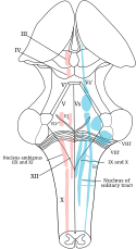
Jądro pasma samotnego
| ||
| Nucleus tractii solitarii vel Nucleus solitarius | ||
 Jądra nerwów czaszkowych - ruchowe (różowe) oraz czuciowe (niebieskie) | ||
| Narządy | Mózgowie | |
Jądro pasma samotnego (łac. nucleus tractus solitarii vel nucleus solitarius) – znajduje się w rdzeniu przedłużonym. Wraz z innymi częściami mózgowia odpowiada za integrację funkcji autonomicznego układu nerwowego, odgrywa też istotną rolę w funkcjonowaniu zmysłu smaku.
Jądro pasma samotnego należy do najstarszych filogenetycznie części mózgowia. Jego obecność stwierdza się u wszystkich ssaków[1], odpowiedniki istnieją również u innych kręgowców.
Budowa
Jądro pasma samotnego stanowi wydłużone skupisko komórek nerwowych występujące na niemal całej długości rdzenia przedłużonego. Poprzez środkową część jądra przebiega szlak samotny, zawierający włókna aferentne z nerwu twarzowego, nerwu językowo-gardłowego oraz nerwu błędnego, które przekazują doń informacje czuciowe. Impulsy nerwowe z jądra pasma samotnego są przekazywane do znacznych obszarów mózgowia i rdzenia przedłużonego tworząc w ten sposób liczne obwody uczestniczące w regulacji układu autonomicznego.
Komórki nerwowe w obrębie jądra pasma samotnego ułożone są w zależności od swojej funkcji:
- część biorąca udział w asocjacji zmysłu smaku zlokalizowana jest w części przedniej („głowowej”)
- część biorąca udział w regulacji układu krążenia, układu oddechowego oraz układu pokarmowego zlokalizowana jest w części tylnej („ogonowej”)
Połączenia aferentne
Zmysł smaku
- część czuciowa nerwu twarzowego
- część czuciowa nerwu językowo-gardłowego
- część czuciowa nerwu błędnego
Czucie trzewne
Nerw błędny przekazuje do jądra pasma samotnego informacje czuciowe zebrane z:
- baroreceptorów zlokalizowanych w kłębku szyjnym oraz w kłębku aortalnym
- chemoreceptorów oraz mechanoreceptorów zlokalizowanych w
Połączenia eferentne
Impulsy z jądra pasma samotnego są przesyłane do
- podwzgórza (jądro przykomorowe)
- ciała migdałowatego (jądra grupy środkowej)
- wzgórza
- innych części mózgowia, w tym tworu siatkowatego
Funkcja
- Integracja odruchów autonomicznych, np. odruchu Bainbridge’a[2][3][4]
- Uczestnictwo w przewodnictwie oraz integracji czucia smaku[5]
- Regulacja łaknienia i masa ciała|masy ciała[6][7]
Przypisy
- ↑ Bret N. Smith, Ping Dou, William D. Barber, F. Dudek. Vagally evoked synaptic currents in the immature rat nucleus tractus solitarii in an intactin vitro preparation.. „Journal of Physiology”. 512.1, s. 149-162, październik 1998. PMID: 9729625.
- ↑ Graziela T. Blanch, André H. Freiria-Oliveira, David Murphy, Renata F. Paulin i inni. Inhibitory mechanism of the nucleus of the solitary tract involved in the control of cardiovascular, dipsogenic, hormonal, and renal responses to hyperosmolality. „American Journal of Physiology-Regulatory, Integrative and Comparative Physiology”. 304 (7), s. R531–R542, 2013. American Physiological Society. DOI: 10.1152/ajpregu.00191.2012. ISSN 0363-6119 (ang.).
- ↑ DA. Ionescu, G. Lugoji, D. Radula. The nucleus of the solitary tract: a review of its anatomy and functions, with emphasis on its role in a putative central-control of brain-capillaries permeability.. „Neurol Psychiatr (Bucur)”. 24 (2). s. 69-85. PMID: 2874607.
- ↑ Cytoglobin and Neuroglobin in the Human Brainstem and Carotid Body. W: C. Di Giulio, S. Zara, M. De Colli, R. Ruffini, A. Porzionato: Neurobiology of Respiration. Springer Netherlands, 2013. DOI: 10.1007/978-94-007-6627-3_9. ISBN 978-94-007-6626-6. (ang.)
- ↑ I. Matsumoto. Gustatory neural pathways revealed by genetic tracing from taste receptor cells.. „Biosci Biotechnol Biochem”. 77 (7), s. 1359-62, 2013. DOI: 10.1271/bbb.130117. PMID: 23832339.
- ↑ Alastair S. Garfield, Christa Patterson, Susanne Skora, Fiona M. Gribble i inni. Neurochemical Characterization of Body Weight-Regulating Leptin Receptor Neurons in the Nucleus of the Solitary Tract. „Endocrinology”. 153 (10), s. 4600–4607, 2012. The Endocrine Society. DOI: 10.1210/en.2012-1282. ISSN 0013-7227 (ang.).
- ↑ Amber L. Alhadeff, Laura E. Rupprecht, Matthew R. Hayes. GLP-1 Neurons in the Nucleus of the Solitary Tract Project Directly to the Ventral Tegmental Area and Nucleus Accumbens to Control for Food Intake. „Endocrinology”. 153 (2), s. 647–658, 2012. The Endocrine Society. DOI: 10.1210/en.2011-1443. ISSN 0013-7227 (ang.).
Bibliografia
- Narkiewicz O., Moryś J.: Neuroanatomia czynnościowa i kliniczna. Warszawa: PZWL, 2003, s. 301. ISBN 83-200-2812-4.
- Adam Bochenek, Michał Reicher: Anatomia człowieka – Tom IV: Układ nerwowy ośrodkowy. PZWL, 2009.
![]() Przeczytaj ostrzeżenie dotyczące informacji medycznych i pokrewnych zamieszczonych w Wikipedii.
Przeczytaj ostrzeżenie dotyczące informacji medycznych i pokrewnych zamieszczonych w Wikipedii.
Media użyte na tej stronie
The Star of Life, medical symbol used on some ambulances.
Star of Life was designed/created by a National Highway Traffic Safety Administration (US Gov) employee and is thus in the public domain.Vectorization of File:Gray696.png. Depicts a posterior view of the brain with the thalamus dissected out and the cerebellum removed. The 4th ventricle is splayed open in the center of the image. Major motor (red) and sensory (blue) nuclei are depicted.
Star of life, blue version. Represents the Rod of Asclepius, with a snake around it, on a 6-branch star shaped as the cross of 3 thick 3:1 rectangles.
Design:
The logo is basically unicolor, most often a slate or medium blue, but this design uses a slightly lighter shade of blue for the outer outline of the cross, and the outlines of the rod and of the snake. The background is transparent (but the star includes a small inner plain white outline). This makes this image usable and visible on any background, including blue. The light shade of color for the outlines makes the form more visible at smaller resolutions, so that the image can easily be used as an icon.
This SVG file was manually created to specify alignments, to use only integers at the core 192x192 size, to get smooth curves on connection points (without any angle), to make a perfect logo centered in a exact square, to use a more precise geometry for the star and to use slate blue color with slightly lighter outlines on the cross, the rod and snake.
Finally, the SVG file is clean and contains no unnecessary XML elements or attributes, CSS styles or transforms that are usually added silently by common SVG editors (like Sodipodi or Inkscape) and that just pollute the final document, so it just needs the core SVG elements for the rendering. This is why its file size is so small.


