Kinetosom

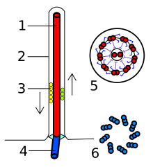
Kinetosom (gr. kinetós = ruchomy, sóma = ciało), ciałko podstawowe – zanurzona w cytoplazmie komórki dolna część pęczku mikrotubul tworzących wici i rzęski. Jest to twór homologiczny do centrioli, odpowiedzialny za ruch wici oraz rzęsek. Koordynację ruchu umożliwia połączenie kinetosomów systemem neurofibryli. W organizmie ludzkim występuje np. w plemnikach.
Bibliografia
- Biologia. Multimedialna encyklopedia PWN Edycja 2.0. pwn.pl, 2008. ISBN 978-83-61492-24-5.
- Encyklopedia Biologia. Agnieszka Nawrot (red.). Kraków: Wydawnictwo GREG. ISBN 978-83-7327-756-4.
Media użyte na tej stronie
Transmission electron microscope image, showing an example of green algae (Chlorophyta).
Chlamydomanas reinhardtii is a unicellular flagellate used as a model system in molecular genetics work and flagellar motility studies.
This image is a longitudinal section through the flagella area. In the cell apex is the basal body that is the anchoring site for a flagella. Basal bodies originate from and have a substructure similar to that of centrioles, with nine peripheral microtubule triplets(see structure at bottom center of image). The two inner microtubules of each triplet in a basal body become the two outer doublets in the flagella. This image also shows the transition region, with its fibers of the stellate structure. The top of the image shows the flagella passing through the cell wall.
Autor: Franciscosp2, Licencja: CC BY-SA 3.0
Eukaryotic flagella. 1-axoneme, 2-cell membrane, 3-IFT (IntraFlagellar Transport), 4-Basal body, 5-Cross section of flagella, 6-Triplets of microtubules of basal body.


