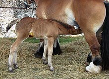
Klacz
Klacz – samica ssaków nieparzystokopytnych[1], w szczególności dorosła samica konia powyżej trzeciego roku życia (potocznie: kobyła).
Cykl płciowy
Okres rujowy u klaczy, zwany grzaniem się, jest sezonowo-wielorujowy. Trwa 5–6 dni, po czym następuje 15 dni przerwy (razem 21 dni cyklu). Owulacja zachodzi w ciągu ostatnich 24–48 godzin rui. Cykl rui może być zróżnicowany, w zależności od wielu czynników: długości dnia, żywienia, czynników atmosferycznych i środowiskowych. Późną jesienią i zimą klacze w ogóle nie miewają rui lub mają ją rzadko, przebiegającą bez objawów. Klacz w rui wykazuje przychylne nastawienie do ogiera podczas próbnikowania; gdy ruja nie występuje, klacz gwałtownie broni się przed jego zalotami. Lekarz weterynarii może pomóc stwierdzić ruję i wskazać, kiedy nastąpi owulacja[2].
Ciąża
Zazwyczaj czas trwania ciąży u klaczy gorącokrwistych waha się od 328 do 345 dni. Natomiast u klaczy zimnokrwistych wynosi od 332 do 345 dni. Zazwyczaj klacz rodzi jedno źrebię. Krycie (sztuczne unasiennianie) powinno być zaplanowane tak, by źrebię urodziło się wiosną[2]. Klacz w ciąży może być bardzo nerwowa i nieufna.
Ciąża bliźniacza
Zdarzają się ciąże bliźniacze, które stanowią ok. 1–2% ciąż. Najczęściej kończą się one poronieniem między 5 a 9 miesiącem ciąży[2]. Około 70% płodów rodzi się martwych, rzadko udaje się odchować jedno źrebię. Lekarz weterynarii może pomóc w zapobieganiu ciąży bliźniaczej poprzez ustalenie ilości i rozmiaru pęcherzyków jajnikowych i wyznaczeniu odpowiedniego terminu krycia[2].
Karmienie
Podczas pierwszych 6 z ok. 11 miesięcy ciąży, klacz powinna być karmiona bez zmian. Bardzo ważne jest natomiast, by zapewnić jej większą dawkę pożywienia podczas 3 ostatnich miesięcy ciąży i uzupełniać paszę składnikami odżywczymi i mikroelementami[2].
Użytkowanie
Podczas ciąży przez około 6 pierwszych miesięcy klacz powinna być użytkowana do lekkiej pracy, by utrzymać jej formę i zapobiec nadwadze, co sprawiałoby kłopoty przy wyźrebieniu. W ciągu ostatnich 3 miesięcy klacz powinna przebywać na pastwisku[2].
Zobacz też
Przypisy
Media użyte na tej stronie
Autor: Sini Merikallio, Licencja: CC BY-SA 2.0
Auringon Loiste serving a mare. Checking if she's fertile.
Très jeune poulain de race ardennaise, en train de têter sa mère.(On voit encore le désinfectant sous le ventre du poulain.)
Autor: Internet Archive Book Images, Licencja: No restrictions
Title: The anatomy of the domestic animals
Identifier: anatomyofdomesti01siss (find matches)
Year: 1914 (1910s)
Authors: Sisson, Septimus, 1865-1924
Subjects: Veterinary anatomy
Publisher: Philadelphia, London, W. B. Saunders Company
Contributing Library: The Library of Congress
Digitizing Sponsor: Sloan Foundation
View Book Page: Book Viewer
About This Book: Catalog Entry
View All Images: All Images From Book
Click here to view book online to see this illustration in context in a browseable online version of this book.
Text Appearing Before Image:
THE FEMALE GENITAL ORGANS The female genital organs (Organa genitalia feminina) are: (1) The two ovaries, the essential reproductive glands, in which the ova are produced; (2) the uterine or Fallopian tubes, which convey the ova to the uterus; (3) the uterus, in which the ovum develops; (4) the vagina, a dilatable passage through which the foetus is expelled from the uterus; (5) the vulva, the terminal segment of the genital tract, which serves also for the expulsion of the urine; (6) the mammary glands, which are in reality glands of the skin, but are so closely associated functionally with the generative organs proper that they are usually described with them. GENITAL ORGANS OF THE MARE THE OVARIES Tlie ovaries (Ovaria) of the mare are hean-shapcd, and are much smaller than the testicles. Their size varies much in different subjects, and they are
Text Appearing After Image:
Fig. 530.—Lateral View of Genital Organs and Adjacent Structures of Mare. It is to be noted that the removal of the other abdominal viscera has allowed the ovaries and uterus to sink down; this has, however, the advantage of showing the broad ligaments of the uterus. 1, Left ovary; 2, uterine or Fallopian tube; S, left cornu uteri; 4. right cornu uteri; 5, corpus uteri; 5', portio vaginalis uteri, and 5", os uteri, seen through window cut in vagina: 6, broad ligament of uterus; 6'", round ligament of uterus; 7, vagina; 5, labia vulvae; 5, rima vulvae; 9\ dorsal commissure, and 9", ventral commissure, of vulva; 10, constrictor vulvae; 11, position of vestibular bulb; IS, ventral wall of abdomen; IS, left kidney; H, left ureter; IB, urinary bladder; 16, urethra; 17, rectum; 18, anus; 19, 19', unpaired and paired parts of sphincter ani externus; 20, retractor ani cut at disappearance under sphincter ani externus; 21, suspensory ligament of anus; 22, longitudinal muscular layer of rectum; 22\ recto-coccygeus; 23, constrictor vaginae; a, utero-ovarian artery, with ovarian (a') and uterine (a") branches; b, uterine artery; c, um- bilical artery; d, ischium; e, pubis; /, ilium, (.\fter Elleuberger, in Leisering's Atlas.) normally larger in young than in old animals; one ovary is often larger than the other. They are about three inches (ca. 7 to 8 cm.) long and an inch to an inch 596
Note About Images


