Konduktor (anatomia)
Konduktor (łac. conductor) – element męskich narządów rozrodczych niektórych pająków.
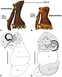
Konduktor zlokalizowany jest, jak reszta aparatu kopulacyjnego pająków, na nogogłaszczkach samca. Narząd ten jest zesklerotyzowaną strukturą położoną w sąsiedztwie embolusa, która sterczy w czasie kopulacji[1]. Klasyfikowany jest w drugiej (wraz z apofizą środkową)[2][3] lub trzeciej (wraz z embolusem) grupie sklerytów bulbusa[4].
Narządy kopulacyjne samic i samców pająków działają na zasadzie zamka i klucza. Rolą konduktora jest „wstępne zaryglowanie” (ang. preliminary locking) aparatu samca w ciele samicy, które umożliwia precyzyjne wprowadzenie embolusa (właściwego kopulatora) w głąb jej ciała. U rodzaju Agelenopsis ciało samicy najpierw jest pobudzane embolusem od zewnątrz, po czym do jamki kopulacyjnej wprowadzany jest konduktor, a na koniec embolus[5][6].
Konduktor może również służyć zablokowaniu dostępu do narządów rozrodczych samicy ewentualnym późniejszym konkurentom[5]. I tak u rodzaju Cybaeus wewnątrz ciała samicy ulega odłamaniu cały konduktor[5][7], a u rodzaju Cyclosa – jego ząbek[5][8]. Funkcją konduktora może być również usunięcie blokady po poprzednim samcu. U Leucauge mariana haczykowaty wyrostek konduktora służy właśnie temu, przy czym nie wygląda na to, by samo wcześniejsze wprowadzenie do samicy konduktora było u tego gatunku niezbędnym warunkiem do wsunięcia w nią embolusa[5].
Zobacz też
Przypisy
- ↑ Marek Żabka: Rząd: pająki — Araneae. W: Zoologia: Stawonogi. T. 2, cz. 1. Szczękoczułkopodobne, skorupiaki. Czesław Błaszak (red. nauk.). Warszawa: Wydawnictwo Naukowe PWN, 2011, s. 88, 349. ISBN 978-83-01-16568-0.
- ↑ M. Ramírez. The morphology and phylogeny of Dionychan spiders (Araneae: Araneomorphae). „Bulletin of the American Museum of Natural History”. 390, s. 1–374, 2014.
- ↑ Rainer F. Foelix: Biology of Spiders. Wyd. III. Oxford University Press, 2011, s. 228. ISBN 978-0-19-973482-5.
- ↑ Michael J. Roberts: Spiders of Britain & Northern Europe. Londyn: HarperCollins, 1995, s. 18.
- ↑ a b c d e W. G. Eberhard, B. A. Huber: 12. Spider GenitaliaP recise Maneuvers with a Numb Structure in a Complex Lock. W: Janet L. Leonard, Alex Córdoba-Aguilar: The evolution of primary sexual characters in animals. Oxford University Press, 2010. ISBN 978-0-19-971703-3.
- ↑ R. L. Gering. Structure and function of the genitalia in some American agelenid spiders. „Smithsonian Miscellaneous Collections”. 121, s. 1–84, 1953.
- ↑ Y. Ihara. Geographic variation and body size differentiation in the medium-sized spe- cies of the genus Cybaeus (Araneae: Cybaeidae) in northern Kyushu, Japan, with descriptions of two new species. „Acta Arachnologica”. 56, s. 1–14, 2007.
- ↑ H. W. Levi. The neotropical and Mexican orb weavers of the genera Cyclosa and Allopecosa (Araneae: Araneidae). „Bulletin of the Museum of Comparative Zoology”. 155, s. 299–379, 199.
Media użyte na tej stronie
Autor: Internet Archive Book Images, Licencja: No restrictions
Identifier: smithsonianmisce1211955smit (find matches)
Title: Smithsonian miscellaneous collections
Year: 1862 (1860s)
Authors: Smithsonian Institution
Subjects: Science
Publisher: Washington : Smithsonian Institution
Contributing Library: Smithsonian Libraries
Digitizing Sponsor: Smithsonian Libraries
View Book Page: Book Viewer
About This Book: Catalog Entry
View All Images: All Images From Book
Click here to view book online to see this illustration in context in a browseable online version of this book.
Text Appearing Before Image:
ribed by Gaubert(1892) in the legs of spiders. Ellis (1944) showed that this platenormally lies in a horizontal position. A special muscle, the levator. Explanation of lettering on figures al alveolus. <7 guide. an anelli. Ih labrum. at atrium. Ip lunate plate. be bursa copulatrix. ma median apophysis. bd blind tube of diverticle. mJi middle haematodocha. bh basal haematodocha. n notch of median apophysis. bs blind tube of spermathecum. P patella. c coxa. pm posterior median sclerite. cc coupling cavity. pt petiole. cd conductor. r reservoir. cl chelicera. rd radix. cm cymbium. sm sternum. cr carinate ridge of chelicera. sp spermathecum. ct connecting tube. St subtegulum. dv diverticle. tb tibia. ed ejaculatory duct. to tubercle. ef epigastric furrow. tg tegulum. em embolus. tgr tegular ridge. en endite of coxa. tm tethering membrane. f femur. tp tibial process. id fundus. tr trochanter. it fertilization tube. u uterus. V vagina. NO. 4 GENITALIA IN SOME AGELENID SPIDERS—GERING II
Text Appearing After Image:
Figs. i-6 I, Pedipalpus, left, ectal aspect, Agelenopsis aperta (Gertsch). 2, Pedipalpus,left, dorsal aspect, A. aperta. 3, Mouth region, subventral aspect, A. aperta. 4,Genital bulb, left, unexpanded, frontal aspect, diagrammatic, genus Agelenopsis.5, Receptaculum seminis, diagrammatic. 6, Genital bulb, left, expanded, subectalaspect, diagrammatic, genus Agelenopsis. (For explanation of lettering see p. 10.) 12 SMITHSONIAN MISCELLANEOUS COLLECTIONS VOL. 121 of the chitinous plate (Ellis, op, cit., p. 47) can draw the plate into avertical position, and thus largely limit the increased haemolymphaticpressure, with its resultant extension, to the femur, trochanter, andcoxa of the leg. The writers observations and dissections indicatethat a chitinous plate plays a similar role in the pedipalpi. Being the longest single segment of the pedipalpus, the femur playsa major role in palpal movements and general orientation of the moredistal genital bulb. Patella.—The patella articulates proxi
Note About Images
Autor: Michael G. Rix and Mark S. Harvey, Western Australia Museum, Licencja: CC BY 3.0
Diagnostic characters of Zephyrarchaea and Austrarchaea. A–B, Cephalothorax, lateral view, showing differences in carapace height and the position of accessory setae on male chelicerae: A, male Austrarchaea harmsi Rix & Harvey; B, male Zephyrarchaea marki C–D, Expanded male pedipalps, retro-ventral view, showing differences in the articulation and fusion of the conductor sclerites: C, Austrarchaea helenae Rix & Harvey; D, Zephyrarchaea marae bH = basal haematodocha; C = conductor; C1–2 = conductor sclerites 1–2; Cy = cymbium; E = embolus; Es = embolic sclerite; H = distal haematodocha; T = tegulum; (TS)1–3 = tegular sclerites 1–3. Scale bars: C–D = 0.2 mm.


