Kręgi piersiowe
Kręgi piersiowe (łac. vertebrae thoracicae, skrót Th) – kręgi należące do odcinka piersiowego kręgosłupa. Kręgi odcinka piersiowego mają powierzchnie stawowe łączące się z żebrami – dołki żebrowe doczaszkowe i doogonowe na trzonach kręgów oraz dołki żebrowe wyrostka poprzecznego. Na wyrostkach poprzecznych ulokowane są wyrostki suteczkowate. Kręgi tego odcinka charakteryzuje krótki trzon oraz spłaszczone głowy i doły. Związane jest to ze słabą ruchomością tego odcinka kręgosłupa. Wyrostki kolczyste przeważnie są wysokie oraz odchylone w tył[1]. Istnieją również kręgi przeciwpochyłe (vertebra anticlinalis) czy też przeponowe (vertebra diaphragmatica), których wyrostki kolczyste nie są odchylone[1]. U człowieka to kręg XI[2], u owcy i psa X, u kozy i świni XII, u bydła XIII, natomiast u konia XVI[3].
W odróżnieniu od kręgów szyjnych liczba kręgów piersiowych u ssaków jest gatunkowo zmienna. U człowieka jest to 12, u psów, kota, bydła, owca, jelenia szlachetnego (Cervus elaphus)[1] oraz kozy 13, u świni 14–15, natomiast u konia 18[1][3].
U ptaków część kręgów piersiowych może być zrośnięta w notarium, można je odnaleźć wśród 5 rzędów ptaków innych, niż wróblowe (stan wiedzy z 1982)[4], odmiennych w swej ekologii; notarium obecne jest również u niektórych wróblowych. Jest to jeden z trzech zrostów kości spotykanych u ptaków, pozostałymi są: synsakrum i pygostyl. Ptasie kręgi piersiowe mogą być zrośnięte tylko w niewielkim stopniu, np. wyłącznie wyrostkami kolczystymi[5].
Galeria
Kręg piersiowy człowieka (widok boczny)

Kręg piersiowy człowieka (widok z góry)

Kręgi piersiowe człowieka

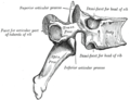
Kręg piersiowy konia. Dołki żebrowe widoczne pod numerem 4.

Kręg piersiowy konia z bardzo dobrze rozwiniętym wyrostkiem kolczystym.

Kręgi piersiowe kota
Przypisy
- ↑ a b c d Simon Hillson: Mammal Bones and Teeth: An Introductory Guide to Methods of Identification. Routledge, 2016, s. 25. ISBN 978-1-315-42500-9.
- ↑ Ronald A. Bergman, Adel K. Afifi, & Ryosuke Miyauchi: Peculiar Thoracic Vertebrae. W: Illustrated Encyclopedia of Human Anatomic Variation: Opus V: Skeletal Systems [on-line]. [dostęp 2018-03-08].
- ↑ a b Helena Przespolewska, Henryk Kobryń, Tomasz Szara & Bartłomiej J. Bartyzel: Podstawy anatomii zwierząt domowych. Warszawa: PWN, 2014, s. 12–14. ISBN 978-83-62815-22-7.
- ↑ Robert W. Storer. Fused Thoracic Vertebrae in Birds. Their Occurrence and Possible Significance. „Journal of the Yamashina Institute for Ornithology”. 14 (2-3), s. 86-95, 1982.
- ↑ James, Helen F.. Repeated Evolution of Fused Thoracic Vertebrae in Songbirds. „The Auk”. 126 (4), s. 862−872, 2009.
![]() Przeczytaj ostrzeżenie dotyczące informacji medycznych i pokrewnych zamieszczonych w Wikipedii.
Przeczytaj ostrzeżenie dotyczące informacji medycznych i pokrewnych zamieszczonych w Wikipedii.
Media użyte na tej stronie

The Star of Life, medical symbol used on some ambulances.
Star of Life was designed/created by a National Highway Traffic Safety Administration (US Gov) employee and is thus in the public domain.
A thoracic vertebra.
_(14769096961).jpg)
Autor: Internet Archive Book Images, Licencja: No restrictions
Dorsal vertebra of horse
Identifier: horseitstreatm05axej (find matches)
Title: The horse, its treatment in health and disease with a complete guide to breeding, training and management
Year: 1906 (1900s)
Authors: Axe, J. Wortley
Subjects: Horses
Publisher: London, Gresham Pub. Co.
Contributing Library: NCSU Libraries
Digitizing Sponsor: NCSU Libraries
View Book Page: Book Viewer
About This Book: Catalog Entry
View All Images: All Images From Book
Click here to view book online to see this illustration in context in a browseable online version of this book.
Text Appearing Before Image:
Fig. 2. ATLAS Untero-inferi Fig. 3. AXIS (side view) 1. Superior spinous process. 4. Odontoid process. 2. Intervertebral foramen. 5. Inferior spinous process. 3. Transverse process. 6. Posterior articular face of body. 7. Oblique process.
Text Appearing After Image:
Fig. 4. DORSAL VERTEBRA (front viI. Superior spinous process. 2. Traiisveprocess. 3. Articulation for tubercle of 14 Articulation for head of rib. 5. Anteiarticular face of body. 6. Spinal c.inal.
Note About Images
_(14749259286).jpg)
Autor: Internet Archive Book Images, Licencja: No restrictions
Dorsal vertebra of horse, front view
Identifier: horseitstreatm05axej (find matches)
Title: The horse, its treatment in health and disease with a complete guide to breeding, training and management
Year: 1906 (1900s)
Authors: Axe, J. Wortley
Subjects: Horses
Publisher: London, Gresham Pub. Co.
Contributing Library: NCSU Libraries
Digitizing Sponsor: NCSU Libraries
View Book Page: Book Viewer
About This Book: Catalog Entry
View All Images: All Images From Book
Click here to view book online to see this illustration in context in a browseable online version of this book.
Text Appearing Before Image:
erior extremity of the body andpasses into the ring of the atlas which is in frontof it. This bone ditiers from the other cervicalvertebrae, in the large size and strength of itssuperior spinous process, the small size of thetransverse processes, and the presence of onlytwo oblique processes, which are behind. The remaining five cervical vertebrae are dis-tinguished numerically as the 3rd, 4th, 5th, 6th.and 7th (fig. 283), and although each possessessome minor distinctive feature, it is not neces-sary to dwell upon them here. The Dorsal Vertebrae (fig. 284) present a good deal in common. Some of them, how-ever, are readily distinguishable from the othersby the length of the superior spinous processes.This is especially the case with regard to thefirst eight bones. Of these the length in-crea.ses to the fifth, and then gradually diminishes backward. The Lumbar Vertebrae (fig. 285) are distinguished from those abovedescribed in the much greater length and width of tlieir transverse pro-
Text Appearing After Image:
Fig. 284.- -Dorsal Vertebra (Front\iew) Superior Spinous Process.- Transverse Process. Articula-tion for Tubercle of Rib. •■ Articu-lation for Head of Rib. 5 ..\ntoriorArticular Face of Body. •* SpinalCanal. 181 HKALTH AM) DISEASE cesses, which are directed horizontally outwards. The last two are muchthicker and somewhat shorter than the rest, and are united to each other
Note About Images

Star of life, blue version. Represents the Rod of Asclepius, with a snake around it, on a 6-branch star shaped as the cross of 3 thick 3:1 rectangles.
Design:
The logo is basically unicolor, most often a slate or medium blue, but this design uses a slightly lighter shade of blue for the outer outline of the cross, and the outlines of the rod and of the snake. The background is transparent (but the star includes a small inner plain white outline). This makes this image usable and visible on any background, including blue. The light shade of color for the outlines makes the form more visible at smaller resolutions, so that the image can easily be used as an icon.
This SVG file was manually created to specify alignments, to use only integers at the core 192x192 size, to get smooth curves on connection points (without any angle), to make a perfect logo centered in a exact square, to use a more precise geometry for the star and to use slate blue color with slightly lighter outlines on the cross, the rod and snake.
Finally, the SVG file is clean and contains no unnecessary XML elements or attributes, CSS styles or transforms that are usually added silently by common SVG editors (like Sodipodi or Inkscape) and that just pollute the final document, so it just needs the core SVG elements for the rendering. This is why its file size is so small.
Thoracic vertebra as seen from top.
_(18167456576).jpg)
Autor: Internet Archive Book Images, Licencja: No restrictions
Title: Anatomy of the cat
Identifier: anatomyofcatrje00reig (find matches)
Year: 1991 (1990s)
Authors: Reighard, Jacob Ellsworth, 1861-1942; Jennings, H. S. (Herbert Spencer), 1868-1947
Subjects: Cats; Mammals
Publisher: (Austin, TX) : BookLab, Inc.
Contributing Library: American Museum of Natural History Library
Digitizing Sponsor: Biodiversity Heritage Library
View Book Page: Book Viewer
About This Book: Catalog Entry
View All Images: All Images From Book
Click here to view book online to see this illustration in context in a browseable online version of this book.
Text Appearing Before Image:
THE VERTEBRAL COLUMN. 5 Differential Characters of the Thoracic Vertebra (Fig. 4). —Following the thoracic vertebrae caudad there is to be seen a gradual increase in the size of the centra brought about by an increase in their craniocaudal and transverse measurements.
Text Appearing After Image:
The dorsoventral measurements remain nearly the same. The costal facets (Fig. 4, c) shift caudad so'that on the eleventh, twelfth, and thirteenth thoracic vertebra; each lies entirely on the cranial end of its centrum, while the caudal end
Note About Images

Autor: Anatomography, Licencja: CC BY-SA 2.1 jp
Thoracic vertebrae (shown in red).