Mikrofotografia
Mikrofotografia – rodzaj fotografii wykonany przy użyciu aparatu fotograficznego połączonego z mikroskopem. Obiekt fotografowany zostaje powiększony za pomocą samego mikroskopu albo obiektywu mikroskopowego zamocowanego przy pomocy nasadki na teleobiektywie.
Mikrofotografowanie jest metodą wykonywania zdjęć obiektów niewidocznych gołym okiem.
Galeria

Aparat szparkowy pomidora
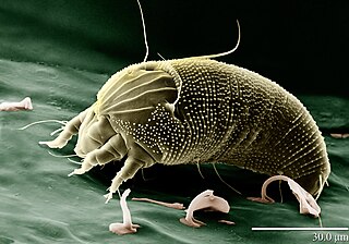
Aceria anthocoptes

Brevipalpus phoenicis
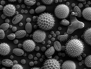
Ziarna pyłku różnych roślin w powiększeniu mikroskopem elektronowym
Zobacz też
Kontrola autorytatywna (gatunek fotograficzny):
Media użyte na tej stronie

Flat mite, Brevipalpus phoenicis.jpg
The Flat Mite (Brevipalpus phoenicis) carries the Leprosis Virus in Citrus, a disease currently in South America but moving North. Magnified 600X. False colors added. (LTSEM) Plate # 27305.
The Flat Mite (Brevipalpus phoenicis) carries the Leprosis Virus in Citrus, a disease currently in South America but moving North. Magnified 600X. False colors added. (LTSEM) Plate # 27305.

E coli at 10000x.jpg
Low-temperature electron micrograph of a cluster of E. coli bacteria, magnified 10,000 times. Each individual bacterium is oblong shaped.
Low-temperature electron micrograph of a cluster of E. coli bacteria, magnified 10,000 times. Each individual bacterium is oblong shaped.

Tomato leaf stomate cropped and scaled.jpg
Cropped and scaled image of a tomato leaf stoma, SEM
Cropped and scaled image of a tomato leaf stoma, SEM

Peacock mite, Tuckerella sp.jpg
The Peacock mite, a beautiful but important pest on citrus in the tropics, shown here on a tea stem. Magnified 260X (with a low-temp scanning electron microscope). (LTSEM) Plate # d27487. Courtesy, Erbe, Pooley: USDA, ARS, EMU. "All of the micrographs on the web site are in the public domain and can be freely used." --Christopher Pooley. Reference: http://emu.arsusda.gov/typesof/default.html English Wikipedia, original upload 23 March 2005 by Brian0918
The Peacock mite, a beautiful but important pest on citrus in the tropics, shown here on a tea stem. Magnified 260X (with a low-temp scanning electron microscope). (LTSEM) Plate # d27487. Courtesy, Erbe, Pooley: USDA, ARS, EMU. "All of the micrographs on the web site are in the public domain and can be freely used." --Christopher Pooley. Reference: http://emu.arsusda.gov/typesof/default.html English Wikipedia, original upload 23 March 2005 by Brian0918

Misc pollen.jpg
Pollen from a variety of common plants: sunflower (Helianthus annuus, small spiky sphericals, colorized pink), morning glory (Ipomoea purpurea, big sphericals with hexagonal cavities, colorized mint green), hollyhock (Sildalcea malviflora, big spiky sphericals, colorized yellow), lily (Lilium auratum, bean shaped, colorized dark green), primrose (Oenothera fruticosa, tripod shaped, colorized red) and castor bean (Ricinus communis, small smooth sphericals, colorized light green). The image is magnified some x500, so the bean shaped grain in the bottom left corner is about 50 μm long.
Pollen from a variety of common plants: sunflower (Helianthus annuus, small spiky sphericals, colorized pink), morning glory (Ipomoea purpurea, big sphericals with hexagonal cavities, colorized mint green), hollyhock (Sildalcea malviflora, big spiky sphericals, colorized yellow), lily (Lilium auratum, bean shaped, colorized dark green), primrose (Oenothera fruticosa, tripod shaped, colorized red) and castor bean (Ricinus communis, small smooth sphericals, colorized light green). The image is magnified some x500, so the bean shaped grain in the bottom left corner is about 50 μm long.

Rust Mite, Aceria anthocoptes.jpg
Electron scan micrography of Aceria anthocoptes.
Electron scan micrography of Aceria anthocoptes.