Zespół Stüve-Wiedemanna

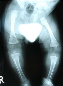
Zespół Stüve-Wiedemanna (Stüve-Wiedemann syndrome, SWS[1], STWS), znany też jako noworodkowa postać zespołu Schwartza-Jampela typu 2 (SJS2) – rzadki zespół wad wrodzonych dziedziczony autosomalnie recesywnie, spowodowany mutacjami w genie LIFR (leukemia inhibitory factor receptor gene) w locus 5p13[2]. Mutacje w genie LIFR prowadzą do niestabilności transkryptu LIFR, zaburzając syntezę białka i szlak sygnalizacyjny JAK/STAT3. Zespół należy do dysplazji kostnych. Na fenotyp SWS składają się:
- niskorosłość,
- łukowate kości długie,
- kamptodaktylia.
Poważnymi powikłaniami są niewydolność oddechowa i niewytłumaczalna hipertermia, będące głównymi przyczynami zgonu dzieci z tym zespołem w okresie noworodkowym[3]. Opisywano rzadkie przypadki dłuższego przeżycia pacjentów z SWS[4][5][6]. Dodatkowo u tych pacjentów obserwowano objawy dysautonomii (zaburzenia smaku, dysfagię)[7].
Chorobę opisali Annemarie Stüve i Hans-Rudolf Wiedemann w 1971 roku[8][9].
Zobacz też
Przypisy
- ↑ Skrót użyty wcześniej wobec zespołu Sturge’a-Webera
- ↑ Dagoneau N, Scheffer D, Huber C, Al-Gazali LI, Di Rocco M, Godard A, Martinovic J, Raas-Rothschild A, Sigaudy S, Unger S, Nicole S, Fontaine B, Taupin JL, Moreau JF, Superti-Furga A, Le Merrer M, Bonaventure J, Munnich A., Legeai-Mallet L, Cormier-Daire V. Null leukemia inhibitory factor receptor (LIFR) mutations in Stuve-Wiedemann/Schwartz-Jampel type 2 syndrome. „American journal of human genetics”. 2 (74), s. 298–305, luty 2004. DOI: 10.1086/381715. PMID: 14740318.
- ↑ Raas-Rothschild A, Ergaz-Schaltiel Z, Bar-Ziv J, Rein AJ. Cardiovascular abnormalities associated with the Stuve-Wiedemann syndrome. „American journal of medical genetics. Part A”. 2 (121A), s. 156–8, sierpień 2003. DOI: 10.1002/ajmg.a.20066. PMID: 12910496.
- ↑ Di Rocco M, Stella G, Bruno C, Doria Lamba L, Bado M, Superti-Furga A. Long-term survival in Stuve-Wiedemann syndrome: a neuro-myo-skeletal disorder with manifestations of dysautonomia. „Am J Med Genet A”. 118A. 4, s. 362-8, 2003. DOI: 10.1002/ajmg.a.10242. PMID: 12687669.
- ↑ Al-Gazali LI, Ravenscroft A, Feng A, Shubbar A, Al-Saggaf A, Haas D. Stüve-Wiedemann syndrome in children surviving infancy: clinical and radiological features. „Clin Dysmorphol”. 12. 1, s. 1-8, 2003. DOI: 10.1097/01.mcd.0000049260.13501.ec. PMID: 12514358.
- ↑ Gaspar IM, Saldanha T, Cabral P, Vilhena MM, Tuna M, Costa C, Dagoneau N, Daire VC, Hennekam RC. Long-term follow-up in Stuve-Wiedemann syndrome: a clinical report. „Am J Med Genet A”. 146A. 13, s. 1748-53, 2008. DOI: 10.1002/ajmg.a.32325. PMID: 18546280.
- ↑ Gardiner J, Barton D, Vanslambrouck JM, Braet F, Hall D, Marc J, Overall R. Defects in tongue papillae and taste sensation indicate a problem with neurotrophic support in various neurological diseases. „Neuroscientist”. 14. 3, s. 240-50, 2008. DOI: 10.1177/1073858407312382. PMID: 18270312.
- ↑ Stüve A, Wiedemann HR. Congenital bowing of the long bones in two sisters. „Lancet”. 2. 7722, s. 495, 1971. PMID: 4105362.
- ↑ Wiedemann HR, Stüve A. Stüve-Wiedemann syndrome: update and historical footnote. „American journal of medical genetics”. 1 (63), s. 12–6, maj 1996. DOI: <12::AID-AJMG5>3.0.CO;2-U 10.1002/(SICI)1096-8628(19960503)63:1<12::AID-AJMG5>3.0.CO;2-U. PMID: 8723080.
Linki zewnętrzne
- STUVE-WIEDEMANN SYNDROME w bazie Online Mendelian Inheritance in Man (ang.)
- Stüve-Wiedemann syndrome w bazie Who Named It (ang.)
![]() Przeczytaj ostrzeżenie dotyczące informacji medycznych i pokrewnych zamieszczonych w Wikipedii.
Przeczytaj ostrzeżenie dotyczące informacji medycznych i pokrewnych zamieszczonych w Wikipedii.
Media użyte na tej stronie
The Star of Life, medical symbol used on some ambulances.
Star of Life was designed/created by a National Highway Traffic Safety Administration (US Gov) employee and is thus in the public domain.Autor: see above, Licencja: CC BY 2.0
"Proband's photo showed expressionless face associated with typical pursed appearance of the mouth, blepharophimosis, multiple contractures and umbilical hernia."
Autor: see above, Licencja: CC BY 2.0
"AAnteroposterior lower limb radiograph showed epiphyseal dysplasia of the capital femoral epiphyses, hypoplastic ileae, horizontally dysplastic acetabulae, coxa vara, shortening of the femoral necks, broad femora and tibiae with bilateral but asymmetrical degrees of mild bowing. In addition the angulation of the femora was associated with internal thickening of the cortex."
Autor: see above, Licencja: CC BY 2.0
"Anteroposterior chest radiograph Chest radiogram showed a narrow upper thoracic cage (Bell-like) but with normal heart borders."


