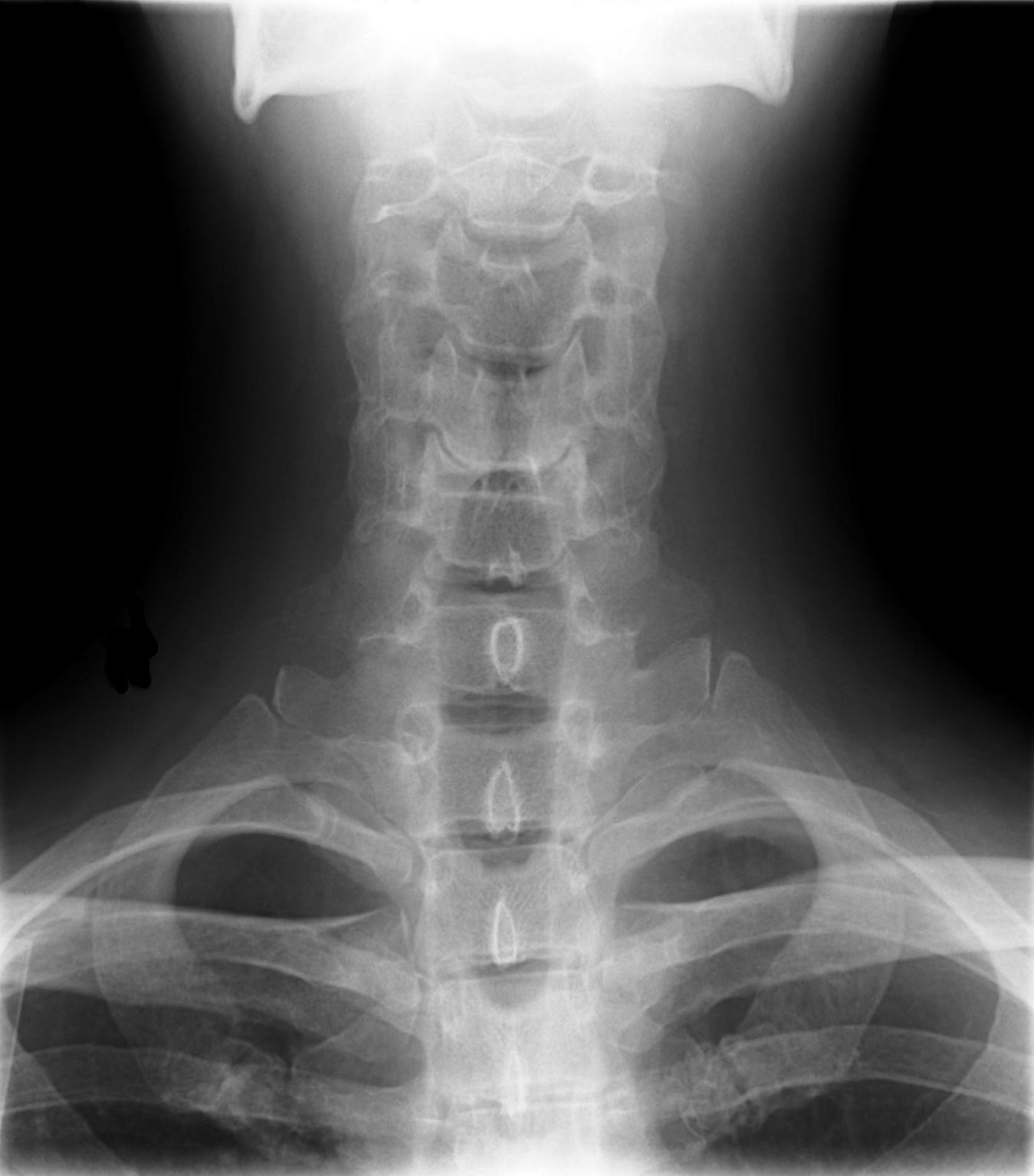
Cervical Xray Lower AP View
Autor:
Credit:
own medical image, work for hire
Krótki link:
źródło:
Wymiary:
1349 x 1534 Pixel (106753 Bytes)
Opis:
X-ray of cervical spine (neck) AP (front) view. This series of x-rays were part of pre-surgical evaluation to help identify spinal instability. Patient is a 37 year old male with a history of multiple neck traumas with pain and muscle spasms and dental implant in lower jaw. Excerpt from radiologist's report:
- FINDINGS: Five views of the cervical spine, including flexion and extension, were performed. There is no evidence of fracture, bone destruction, or malalignment. There are degenerative bone and is changes at C5-6. There is no evidence of cervical instability on the flexion and extension views. The facet joints are well aligned. Bony spurring is narrowing the C5-6 neural foramina bilaterally.
- IMPRESSION: Degenerative changes at C5-6. No evidence of instability.
Licencja:
Warunki licencji:
Creative Commons Zero, Public Domain Dedication
Więcej informacji o licencji można znaleźć tutaj. Ostatnia aktualizacja: Wed, 12 Oct 2022 23:05:50 GMT