Coarctation and PDA
Autor:
Autor nie został podany w rozpoznawalny automatycznie sposób. Założono, że to Ekko (w oparciu o szablon praw autorskich).
Credit:
Źródło nie zostało podane w rozpoznawalny automatycznie sposób. Założono, że to praca własna (w oparciu o szablon praw autorskich).
Krótki link:
źródło:
Wymiary:
555 x 568 Pixel (44417 Bytes)
Opis:
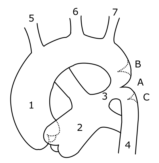
Schematic drawing of alternative locations of a coarctation of the aorta. I, Kjetil Lenes, have made the drawing myself, after information from Valdes-Cruz LM, Cayre RO: Echocardiographic diagnosis of congenital heart disease. Philadelhia, 1998.. Legend: A: ductal coarctation, B: preductal coarctation, C: postductal coarctation. 1: Aorta ascendens, 2: Arteria pulmonalis, 3: Ductus arteriosus, 4: Aorta descendens, 5: Trunchus brachiocephalicus, 6: Arteria carotis communis sinister, 7: Arteria subclavia sinister
The picture is somewhat misleading, with left pulmonary artery crossing behind aorta. This will be changed in a future drawing.
Licencja:
Public domain
Więcej informacji o licencji można znaleźć tutaj. Ostatnia aktualizacja: Mon, 07 Nov 2022 03:00:14 GMT