HLA-A1
Autor:
Credit:
Praca własna
Krótki link:
źródło:
Wymiary:
548 x 609 Pixel (148929 Bytes)
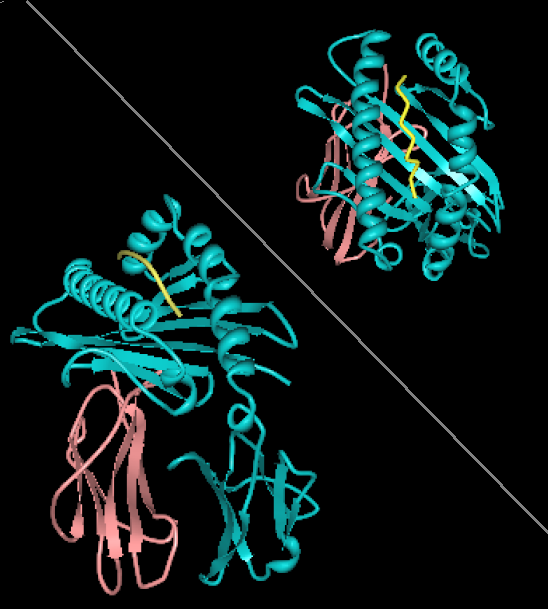
Opis:
Rendering of HLA-A1 with MAGE-1 bound peptide. Two views, from the side showing B2 microglobulin (rose) and HLA-A1 (alpha chain, cyan). Top-right view is looking down through the binding site toward the plasma membrane. The image is derived from PDB:1W72 that was presented in the work:Hulsmeyer et al. (2005) A major histocompatibility complex-peptide-restricted antibody and t cell receptor molecules recognize their target by distinct binding modes: crystal structure of human leukocyte antigen (HLA)-A1-MAGE-A1 in complex with FAB-HYB3. J.Biol.Chem. 280: 2972-2980. Image rendered with PDB ProteinWorkshop 1.50. Several aspects of the image removed for clarity reasons.
Licencja:
Public domain
Więcej informacji o licencji można znaleźć tutaj. Ostatnia aktualizacja: Sun, 11 Dec 2022 03:04:01 GMT