Mixed Tumor of the Salivary Gland
Mixed Tumor of the Salivary Gland
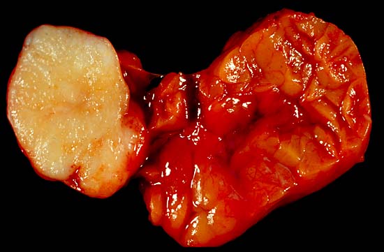
This benign tumor of the submandibular gland, also known as pleomorphic adenoma, presented as a painless neck mass in a 40-year-old man. At the left of the image is the white tumor with its characteristic cartilaginous cut surface. To the right is the normally lobated submandibular salivary gland.
Unlike most of my gross photos, this one was shot in the fresh state. This does show best what tissue looks like to the operating surgeon, but the soft, collapse-prone tissue, with its blood staining and distracting highlights, doesn't show anatomic details as well as do photos of formalin-fixed specimens. This pic was shot with a Minolta X-370 with a 100-mm Rokkor bellows lens, on Kodak Elite daylight ISO 100 film, using a blue compensator to correct for tungsten illumination.
Photograph by Ed Uthman, MD. Public domain. Posted 13 May 00Więcej informacji o licencji można znaleźć tutaj. Ostatnia aktualizacja: Thu, 24 Mar 2022 20:43:52 GMT