Nicolau syndrome (livedoid dermatitis) Macroscopic view, microscopic view and particles analysis of a representative cutaneous lesion obtained from the same patient
Autor:
Régis Bouquié, Laura Wainstein, Paul Pilet, Jean-Marie Mussini, Guillaume Deslandes, Johann Clouet, Eric Dailly, Pascale Jolliet, Caroline Victorri-Vigneau
Attribution:
Obraz jest oznaczony jako „Wymagane uznanie autorstwa” (attribution required), ale nie podano żadnych informacji o uznaniu autorstwa. Prawdopodobnie parametr atrybucji został pominięty podczas korzystania z szablonu MediaWiki dla licencji CC-BY. Autorzy mogą znaleźć tutaj przykład prawidłowego korzystania z szablonów.
Credit:
Crushed and injected buprenorphine tablets: characteristics of princeps and generic solutions. PLoS One. 2014; 9(12): e113991]
Krótki link:
źródło:
_Macroscopic_view,_microscopic_view_and_particles_analysis_of_a_representative_cutaneous_lesion_obtained_from_the_same_patient.png?uselang=pl)
Wymiary:
512 x 462 Pixel (386667 Bytes)
Opis:
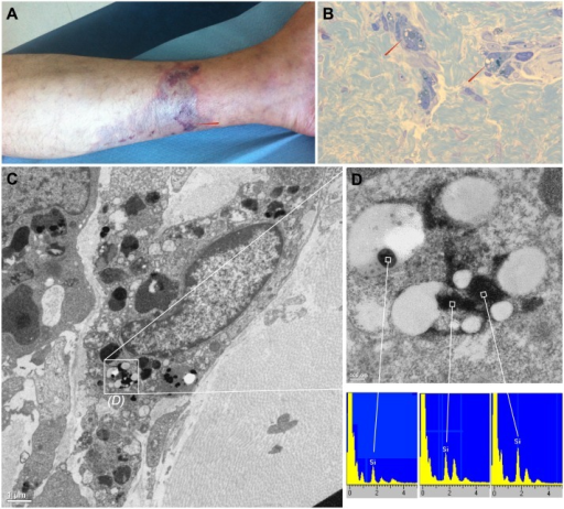
Macroscopic view, microscopic view and particles analysis of a representative cutaneous lesion obtained from the same patient.A: necrotic livedo-like dermatitis lesion from the lower left leg. The red arrow represents the injection site. B: macrophages easily distinguished by CD68 immunolabelling among a perivascular inflammatory infiltrate exhibited few refringent material (red arrows). C: transmission electron microscopy, we have observed non organic very dense particles, without epoxy permeation but with either a round shape compatible with silica or with a laminated aspect compatible with silicate. D: High magnification of figure 1C inlay by transmission electron microscopy and energy-dispersive x-ray spectroscopy spectra analysis. Particles primarily identified as very dense and non organic contain silica (Si).
Licencja:
Warunki licencji:
Creative Commons Attribution 4.0
Więcej informacji o licencji można znaleźć tutaj. Ostatnia aktualizacja: Mon, 17 May 2021 18:20:47 GMT