Oncocytoma of the Salivary Gland
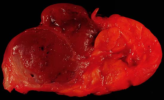
Oncocytoma of the Salivary Gland
This lesion presented as a lateral anterior neck mass. At surgery, it was found to be a soft 3.0 x 2.1 x 1.8 cm tumor of the submandibular salivary gland. The photo shows the characteristic dark color of an oncocytoma, a rare type of benign neoplasm, at the left side of the image (the normal lobulated salivary gland tissue is to the right). Excision is curative.
Since this specimen was photographed in the fresh state, it is various shades of red due to blood staining. A little formalin fixation would be expected to better emphasize the color difference between the tumor and the normal gland tissue. The photo was shot with a Minolta X-370 with 100mm Rokkor bellows lens on Kodak Elite ISO 100 daylight-balanced transparency film. I used a blue filter to compensate for the tungsten illumination.
Photograph by Ed Uthman, MD. Public domain. Posted 19 May 00Więcej informacji o licencji można znaleźć tutaj. Ostatnia aktualizacja: Fri, 13 Jan 2023 06:32:42 GMT