Right bundle branch block ECG characteristics
Autor:
Attribution:
Obraz jest oznaczony jako „Wymagane uznanie autorstwa” (attribution required), ale nie podano żadnych informacji o uznaniu autorstwa. Prawdopodobnie parametr atrybucji został pominięty podczas korzystania z szablonu MediaWiki dla licencji CC-BY. Autorzy mogą znaleźć tutaj przykład prawidłowego korzystania z szablonów.
Credit:
I drew this image in Xara X¹ using my own knowledge and several sources for checking whether I drew the image correctly.
Krótki link:
źródło:
Wymiary:
218 x 200 Pixel (10175 Bytes)
Opis:
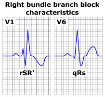
The characteristic wave patterns of a typical right bundle branch block as seen in an ECG. Only the precordial lead V1 and V6 are shown. Wide QRS complexes are present and there's T wave inversion in lead V1 which is normal in this condition. Note the typical wide and deep s wave in V6. The small q wave in V6 may not always be present. Below each QRS complex is its designation (rSR and qRs) according to nomenclature.
Licencja:
Komentarz do licencji:
Ja, właściciel praw autorskich do tego dzieła, udostępniam je na poniższej licencji
Warunki licencji:
Creative Commons Attribution-Share Alike 3.0
Więcej informacji o licencji można znaleźć tutaj. Ostatnia aktualizacja: Mon, 21 Nov 2022 21:42:18 GMT