Sinoatrial node low mag

Autor:
Attribution:
Obraz jest oznaczony jako „Wymagane uznanie autorstwa” (attribution required), ale nie podano żadnych informacji o uznaniu autorstwa. Prawdopodobnie parametr atrybucji został pominięty podczas korzystania z szablonu MediaWiki dla licencji CC-BY. Autorzy mogą znaleźć tutaj przykład prawidłowego korzystania z szablonów.
Credit:
Praca własna
Krótki link:
źródło:
Wymiary:
2848 x 4272 Pixel (3596308 Bytes)
Opis:
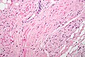
Micrograph of the sinoatrial (SA) node. H&E stain.
The SA node fibre vaguely resemble cardiac myocytes; however, they are thinner, squiggly and stain less intensely (on H&E) than cardiac myocytes.
Description of the micrograph - the follow things are seen:
- Cardiac myocytes of the right atrium are seen at the right/bottom.
- Nodal artery, a branch of the right coronary artery is seen in the centre - on lumen.
- Sinoatrial node surrounds the nodal artery and abuts the darker staining cardiac myocytes.
- Lumen of the right atrium is on the left (endothelial lining not seen).
- Epicardial apidose tissue is to the right of the SA node.
- Epicardial space is on the right of the image.
- Superior vena cava is at the top of the image.
- Adjacent to nodal tissue is a nerve; the SA node interacts with fibres from the vagus nerve.
Related images
High mag. image of the same SA node.

Low mag. image of the same SA node.


Licencja:
Warunki licencji:
Creative Commons Attribution-Share Alike 3.0
Więcej informacji o licencji można znaleźć tutaj. Ostatnia aktualizacja: Sun, 14 Aug 2022 09:46:47 GMT