Zwyrodnienie Wallera

Zwyrodnienie Wallera (pierwotne zwyrodnienie aksonalne, ang. Wallerian degeneration) – zmiana neuropatologiczna, występująca wskutek przerwania ciągłości włókna nerwowego. Towarzyszy przecięciom nerwu, zmiażdżeniom, nadmiernym rozciągnięciom, niedokrwieniu nerwu. Nazwa upamiętnia odkrywcę zjawiska, Augustusa Wallera, który zmiany tego typu opisał u żab z przeciętym nerwem podjęzykowym i językowo-gardłowym w 1850 roku[1].
Przerwanie ciągłości włókna nerwowego powoduje zmiany w aksonie i w otoczce mielinowej. Dalsza, odcięta część aksonu ulega degeneracji w pierwszej kolejności. Mikroskopowo obserwuje się dezintegrację cytoszkieletu i akumulację organelli. Następnie dochodzi do zaburzeń osłonki mielinowej: powstają tzw. komory niedokrwienne, owoidy, złogi lipidowe, naciek makrofagów. Puste, pozbawione aksonów osłonki mielinowe, mogą przetrwać do 52 dni.
Przypisy
- ↑ Waller A: Experiments on the section of glossopharyngeal and hypoglossal nerves of the frog and observations of the alternatives produced thereby in the structure of their primitive fibers. Philos Trans R Soc Lond Biol 1850, 140:423.
Bibliografia
- Paweł P Liberski, Wielisław Papierz, Wojciech Kozubski, Iwona Kłoszewska, Mirosław Jan Mossakowski: Neuropatologia Mossakowskiego. Lublin: Wydawnictwo Czelej, 2005, s. 21, 856-857. ISBN 83-89309-63-7.
![]() Przeczytaj ostrzeżenie dotyczące informacji medycznych i pokrewnych zamieszczonych w Wikipedii.
Przeczytaj ostrzeżenie dotyczące informacji medycznych i pokrewnych zamieszczonych w Wikipedii.
Media użyte na tej stronie
The Star of Life, medical symbol used on some ambulances.
Star of Life was designed/created by a National Highway Traffic Safety Administration (US Gov) employee and is thus in the public domain.Autor: see above, Licencja: CC BY 2.0
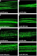
After a latency period Wallerian degeneration following cut and crush injury starts abruptly in single axons and involves total fragmentation of axons within few hours A-D: Conventional fluorescence micrographs of a ~2.5 cm long peripheral nerve stump (sciatic-tibial nerve segment) wholemount preparation at the proximal (A) and distal site (B) 37 h after cut injury with few individual fluorescent axons broken into fragments. A small number of axons fragmented at the proximal (C) and distal site (D) of a peripheral nerve stump wholemount preparation could also be detected 40 h following crush injury. E-H: Conventional fluorescence micrographs of a ~2.5 cm long peripheral nerve stump (sciatic-tibial nerve segment) wholemount preparation at the proximal (E) and distal site (F) 42 h after cut injury with most YFP labelled axons fragmented. A similar picture with a majority of axons degenerated is evident at the proximal (G) and distal end (H) of a peripheral nerve stump wholemount preparation 44 h after crush injury. YFP fluorescence has been pseudo-coloured green with the applied imaging software (MetaVue, Universal Imaging Corporation). Magnification: 100 ×

